


中島君、片山さん、白石くんの3人の学生や、多くのOffice Assistantの協力を得て、6年かけて解析した論文「ヒト胚子期における脳形態形成」がNeuroimageに受諾されました。また、SupplyのビデオはData in Briefに、3D元データの一部はMorphoMに掲載されました。
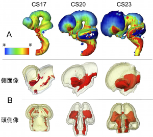
- ヒト胚子期の脳の三次元形成を提示
- 脳の容積は、CS 13 – 23 間で 164.4 倍に増加
- 胚全体に対する脳実質の体積の比率は、CS15-23の間でほぼ一定(11.6〜15.5%)
- 分化成長にともなう脳組織の不均一な厚さを 3D で視覚化
- 結果は、胚子期の脳の発生を定量化し評価する基準値になりうる可能性
13. Shiraishi N, Katayama A, Nakashima T, Yamada S, Uwabe C, Kose K, Takakuwa T, Morphology and morphometry of the human embryonic brain: A three-dimensional analysis, NeuroImage, 2015, 115, 96-103, 10.1016/j.neuroimage.2015.04.044, (概要), [OpenAccess]
12. Shiraishi N, Katayama A, Nakashima T, Yamada S, Uwabe C, Kose K, Takakuwa T, Three-dimensional morphology of the human embryonic brain, Data in Brief, 2015, 4, 116-118, 10.1016/j.dib.2015.05.001 [OpenAccess]
15. Shiraishi N, Katayama A, Nakashima T, Shiraki N, Yamada S, Uwabe C, Kose K, Takakuwa T, 3D model related to the publication: Morphology of the human embryonic brain and ventricles, MorphoMuseuM 1 (3)-e3. doi: 10.18563/m3.1.3.e3. [OpenAccess]
Abstract
The three-dimensional dynamics and morphology of the human embryonic brain have not been previously analyzed using modern imaging techniques. The morphogenesis of the cerebral vesicles and ventricles was analyzed using images derived from human embryo specimens from the Kyoto Collection, which were acquired with a magnetic resonance microscope equipped with a 2.35-T superconducting magnet. A total of 101 embryos between Carnegie stages (CS) 13 and 23, without apparent morphological damage or torsion in the brain ventricles and axes, were studied. To estimate the uneven development of the cerebral vesicles, the volumes of the whole embryo and brain, prosencephalon, mesencephalon, and rhombencephalon with their respective ventricles were measured using image analyzing Amira™ software. The brain volume, excluding the ventricles (brain tissue), was 1.15 ± 0.43 mm3 (mean ± SD) at CS13 and increased exponentially to 189.10 ± 36.91 mm3 at CS23, a 164.4-fold increase, which is consistent with the observed morphological changes. The mean volume of the prosencephalon was 0.26 ± 0.15 mm3 at CS13. The volume increased exponentially until CS23, when it reached 110.99 ± 27.58 mm3. The mean volumes of the mesencephalon and rhombencephalon were 0.20 ± 0.07 mm3 and 0.69 ± 0.23 mm3 at CS13, respectively; the volumes reached 21.86 ± 3.30 mm3 and 56.45 ± 7.64 mm3 at CS23, respectively. The ratio of the cerebellum to the rhombencephalon was approximately 7.2% at CS20, and increased to 12.8% at CS23. The ratio of the volume of the cerebral vesicles to that of the whole embryo remained nearly constant between CS15 and CS23 (11.6–15.5%). The non-uniform thickness of the brain tissue during development, which may indicate the differentiation of the brain, was visualized with surface color mapping by thickness. At CS23, the basal regions of the prosencephalon and rhombencephalon were thicker than the corresponding dorsal regions. The brain was further studied by the serial digital subtraction of layers of tissue from both the external and internal surfaces to visualize the core region (COR) of the thickening brain tissue. The COR, associated with the development of nuclei, became apparent after CS16; this was particularly visible in the prosencephalon. The anatomical positions of the COR were mostly consistent with the formation of the basal ganglia, thalamus, and pyramidal tract. This was confirmed through comparisons with serial histological sections of the human embryonic brain. The approach used in this study may be suitable as a convenient alternative method for estimating the development and differentiation of the neural ganglia and tracts. These findings contribute to a better understanding of brain and cerebral ventricle development.
Highlights
Three-dimensional morphogenesis of the human embryonic brain was presented from MRI.•
The volume of three brain vesicles and ventricles were measured at 101 embryos.•
The brain volume exponentially increased 164.4-fold from Carnegie stage 13 to 23.•
The volume ratio of the brain to whole embryo remained nearly constant.•
The non-uniform thickness of the brain tissue during development was 3D visualized.