


遠藤さんの卒業論文「ヒト胚子脾臓の形態形成」がAnat Recに掲載されました。
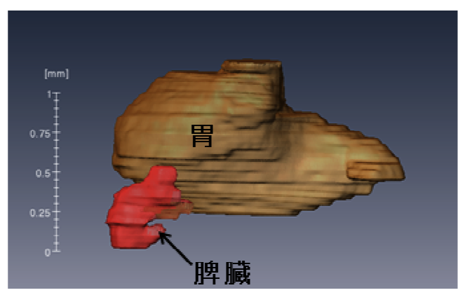
脾臓は、左上腹部の胃の後方に位置する腹腔内の代表的な器官です。脾臓は成人では。成人の脾臓の主な役割は、血液の濾過・不要な物質の除去と免疫系としてのリンパ球の産生です。ヒトにおける初期発生過程はこれまでほとんど知られていませんでした。本研究では、脾臓の形態形成、および脾臓内外の血管の形成過程についてCarnegie stageごとに記載しました。
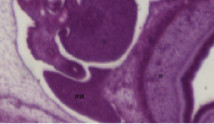
- CS14-17 ;脾臓はdorsal mesogastrium (DM) の膨らみとして認識
- CS16まで;中皮は偽重層、のちに円柱上皮に置換
- CS17以降;基底膜が明瞭、間葉細胞の分化
- CS 18;細胞密度の高い領域が認識、造血細胞の検出
- CS 20 の後;類洞、脾門部の形成
- CS23;動静脈の確認
- 成長速度(長さ)は胃とほぼ同じ
11.Endo A, Ueno S, Yamada S, Uwabe C, Takakuwa T, Morphogenesis of the spleen during the human embryonic period, Anatomical Rec, 2015, 298, 820-826, doi: 10.1002/ar.23099
ABSTRACT
We aimed to observe morphological changes in the spleen from the emergence of the primordium to the end of the embryonic period using histological serial sections of 228 samples. Between Carnegie stages (CSs) 14 and 17, the spleen was usually recognized as a bulge in the dorsal mesogastrium (DM), and after CS 20, the spleen became apparent. Intrasplenic folds were observed later. A high-density area was first recognized in 6 of the 58 cases at CS 16 and in all cases examined after CS 18. The spleen was recognized neither as a bulge nor as a high-density area at CS 13. The mesothelium was pseudostratified until CS 16 and was replaced with high columnar cells and then with low columnar cells. The basement membrane was obvious after CS 17. The mesenchymal cells differentiated from cells in the DM, and sinus formation started at CS 20. Hematopoietic cells were detected after CS 18. The vessels were observed at CS 14 in the DM. Hilus formation was observed after CS 20. The parallel entries of the arteries and veins were observed at CS 23. The rate of increase in spleen length in relation to that of stomach length along the cranial-caudal direction was 0.51 ± 0.11, which remained constant during CSs 19 and 23, indicating that their growths were similar. These data may help to better understand the development of normal human embryos and to detect abnormal embryos in the early stages of development.