


鈴木さんの修士論文がPLoS ONEに受諾されました。
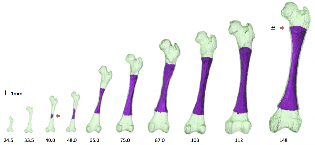
軟骨形成から軟骨内骨化の進む時期の大腿骨の形態形成について外観、内部の変化を位相CT, MRIを用いて解析しました。
- 軟骨性大腿骨は、CS 18 で初めて観察される
- 主要な解剖学的ランドマークは、骨化が開始する前(CRL<40mm)に形成
- 信号強度が高い骨化の開始部位は、しだいに骨幹端軟骨板になる可能性がある骨幹端に限定される
- 骨化部位の長さ /大腿骨の全長はCRL 40 -75 mmで急速に増加し、CRL が 75 mm で中程度に増加。
- 軟骨管は、遠位骨端(CRL、75 mm)よりも近位骨端(CRL、62 mm)で早期に発生
- 骨化後の大腿骨形状の変化は、初期骨化時とその前後に限定的
- ただし、大腿骨頸部の前傾および大腿骨頭のねじれは、胎児期に連続的に変化
Suzuki Y, Matsubayashi J, Ji X, Yamada S, Yoneyama A, Imai H, Matsuda T, Aoyama T, Takakuwa T Morphogenesis of the femur at different stages of normal human development, PLoS ONE, 14(8): e0221569. https://doi.org/10.1371/journal. pone.0221569
Abstract
The present study aimed to better characterize the morphogenesis of the femur from the embryonic to the early fetal periods. Sixty-two human fetal specimens (crown–rump length [CRL] range: 11.4–185 mm) from the Kyoto Collection were used for this study. The morphogenesis and internal differentiation process of the femur were analyzed in 3D using phase-contrast X-ray computed tomography and magnetic resonance imaging. The cartilaginous femur was first observed at Carnegie stage 18. Major anatomical landmarks were formed prior to the initiation of ossification at the center of the diaphysis (CRL, 40 mm), as described by Bardeen. The region with very high signal intensity (phase 5 according to Streeter’s classification; i.e., area described as cartilage disintegration) emerged at the center of the diaphysis, which split the region with slightly low signal intensity (phase 4; i.e., cartilage cells of maximum size) in fetuses with a CRL of 40.0 mm. The phase 4 and phase 5 regions became confined to the metaphysis, which might become the epiphyseal cartilage plate. Femur length and ossified shaft length (OSL) showed a strong positive correlation with CRL. The OSL-to-femur length ratio rapidly increased in fetuses with CRL between 40 and 75 mm, which became moderately increased in fetuses with a CRL of ≥75 mm. Cartilage canal invasion occurred earlier at the proximal epiphysis (CRL, 62 mm) than at the distal epiphysis (CRL, 75 mm). Morphometry and Procrustes analysis indicated that changes in the femur shape after ossification were limited, which were mainly detected at the time of initial ossification and shortly after that. In contrast, femoral neck anteversion and torsion of the femoral head continuously changed during the fetal period. Our data could aid in understanding the morphogenesis of the femur and in differentiating normal and abnormal development during the early fetal period.