昼ごはんです
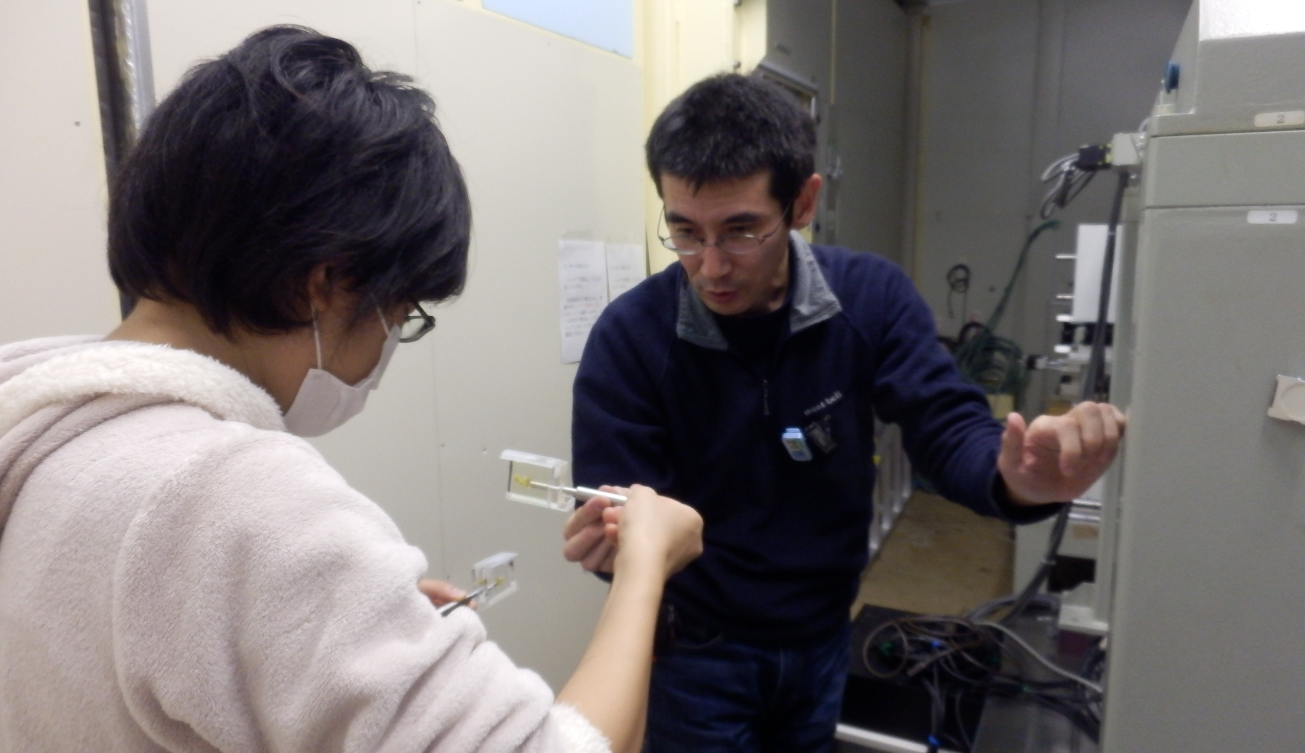
サンプル交換
12/5-8、および12/13-14にPhoton Factory (つくば)で位相X線CTを用いて胚子撮像を行いました。今年度の撮像はこれで終了です。得られた画像は、胚子の詳細な内部観察に使用します。
|
||||
  |
昼ごはんです サンプル交換 12/5-8、および12/13-14にPhoton Factory (つくば)で位相X線CTを用いて胚子撮像を行いました。今年度の撮像はこれで終了です。得られた画像は、胚子の詳細な内部観察に使用します。 発達神経科学会第3回学術集会で発表しました。(2014.10.16-17; 東京) ヒト胚子における Willis 動脈輪の形成について 小池哲平、山田重人、上部千賀子、高桑徹也 ヒト胚子期における脳形態形成の解析 白石直樹、片山愛里、中島 崇、上部千賀子、巨瀬 勝美、山田 重人、高桑 徹也 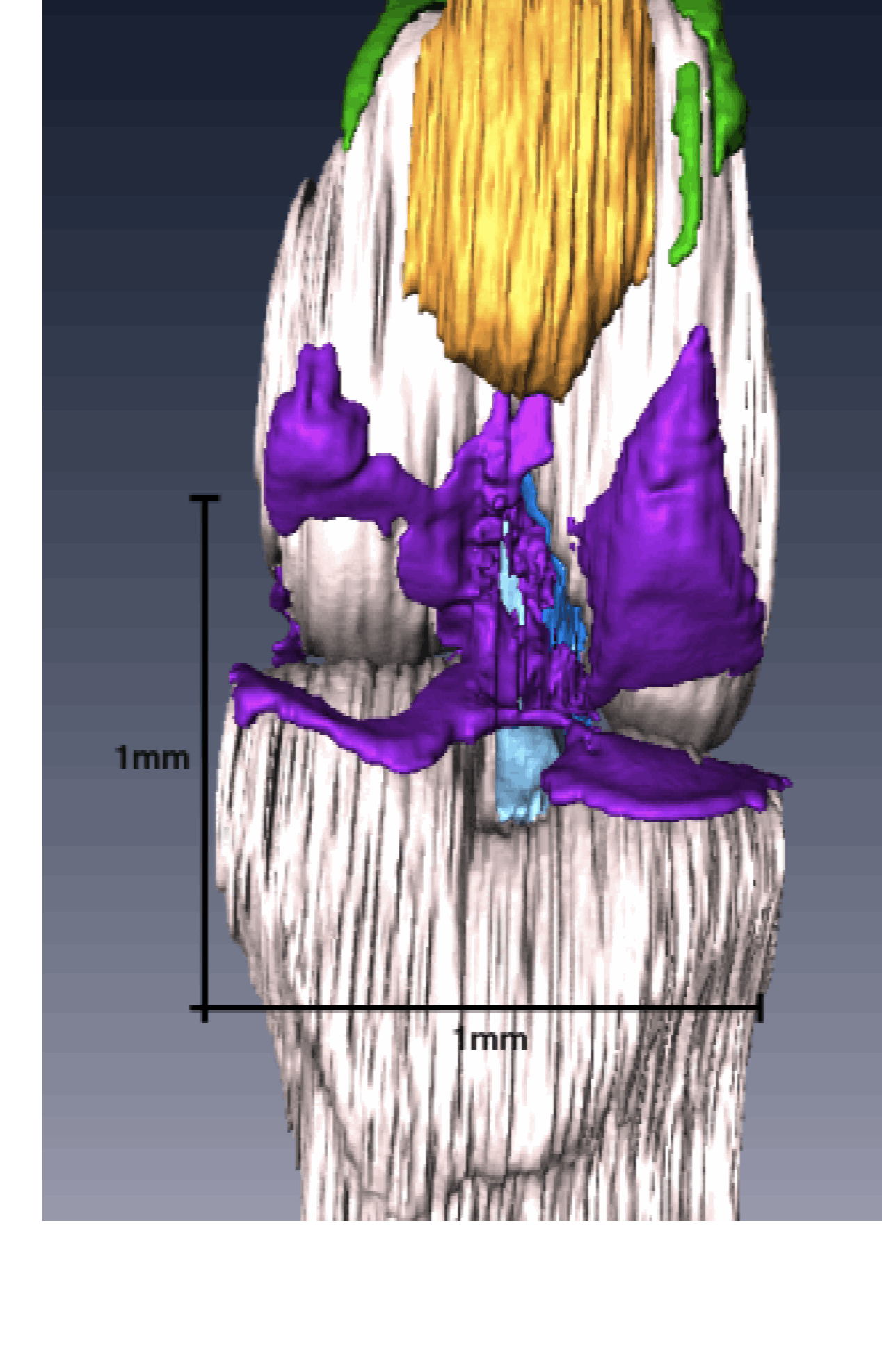 高石くんの卒業論文「膝関節の形態形成; EFICを用いた3次元的解析」がOsteoarthritis Cartilage ( 特集号Imaging in Osteoarthritis)に掲載されました。 9. Takaishi R, Aoyama T, Takakuwa T, et al; Three-dimensional reconstruction of rat knee joint using episcopic fluorescence image capture, Osteoarthritis Cartilage, 2014; 22(10), 1401-1409. 10.1016/j.joca.2014.06.016
SummaryObjectiveDevelopment of the knee joint was morphologically investigated, and the process of cavitation was analyzed by using episcopic fluorescence image capture (EFIC) to create spatial and temporal three-dimensional (3D) reconstructions. MethodsKnee joints of Wister rat embryos between embryonic day (E)14 and E20 were investigated. Samples were sectioned and visualized using an EFIC. Then, two-dimensional image stacks were reconstructed using OsiriX software, and 3D reconstructions were generated using Amira software. ResultsCavitations of the knee joint were constructed from five divided portions. Cavity formation initiated at multiple sites at E17; among them, the femoropatellar cavity (FPC) was the first. Cavitations of the medial side preceded those of the lateral side. Each cavity connected at E20 when cavitations around the anterior cruciate ligament (ACL) and posterior cruciate ligament (PCL) were completed. ConclusionCavity formation initiated from six portions. In each portion, development proceeded asymmetrically. These results concerning anatomical development of the knee joint using EFIC contribute to a better understanding of the structural feature of the knee joint.  Maenner先生と discussion Blechschmidt collectionの標本を研究で使用させていただくため、 ドイツ、ゲッチンゲン大学を訪問致しました。(9/20-27) 今回は、大変貴重な咽頭胚期に相当する胚子の連続組織標本を、フラットスキャナを用いて画像取得をするために訪問致しました。Blechschmidtは胚子の3次元モデル群を保有することでも有名です。今回、3次元スキャナを用いてこのモデルからの立体像取得を試みました。われわれの研究対象であるヒト胚子標本は、貴重な人類共通財産といえます。今後、現存する標本の永久保存、管理、利用を可能にする方法を考えていきたいと考えます。 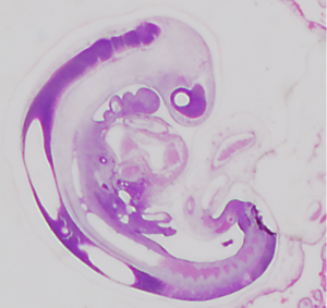 CS12 組織像 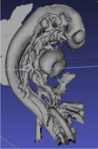 3DスキャンしたCS12胚子モデル 22. Ueno S, Yamada S, Uwabe C, Männer J, Shiraki N, Takakuwa T, The digestive tract and derived primordia differentiate by following a precise timeline in human embryos between Carnegie stages 11 and 13, Anatomical Rec 2016, 299, 439-449, DOI: 10.1002/ar.23314s, (概要)
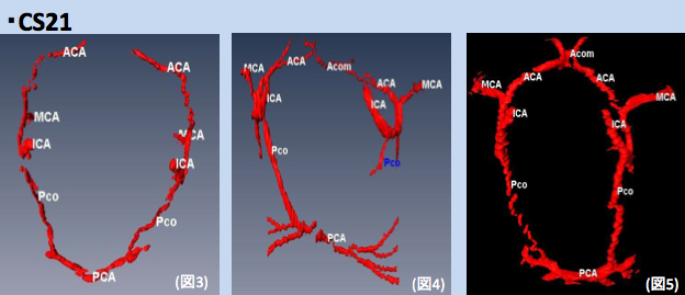 CS21; Circles of Willis circle 個体差がみられる。 第54回日本先天異常学会学術集会で発表しました。 (相模原市、麻布大学、7/26-27) ヒト外耳初期発生過程の解析; 尾関 舞美, 山田 重人, 上部千賀子, 石津 浩一, 高桑 徹也 ヒト消化管の初期形態形成と分化; 上野 沙季, 山田 重人, 高桑 徹也 3 次元プリンタを用いたヒト胚子由来立体模型の作製; 大坂 美穂, 酒井 晃二, 山田 重人,高桑 徹也 ヒト胚子期における脳形態形成の解析; 白石 直樹, 片山 愛里, 中島 崇, 山田 重人, 上部千賀子, 巨瀬 勝美, 高桑 徹也 昨年秋、YouTubeにHomePageを開設しました。(‘13,11,20)
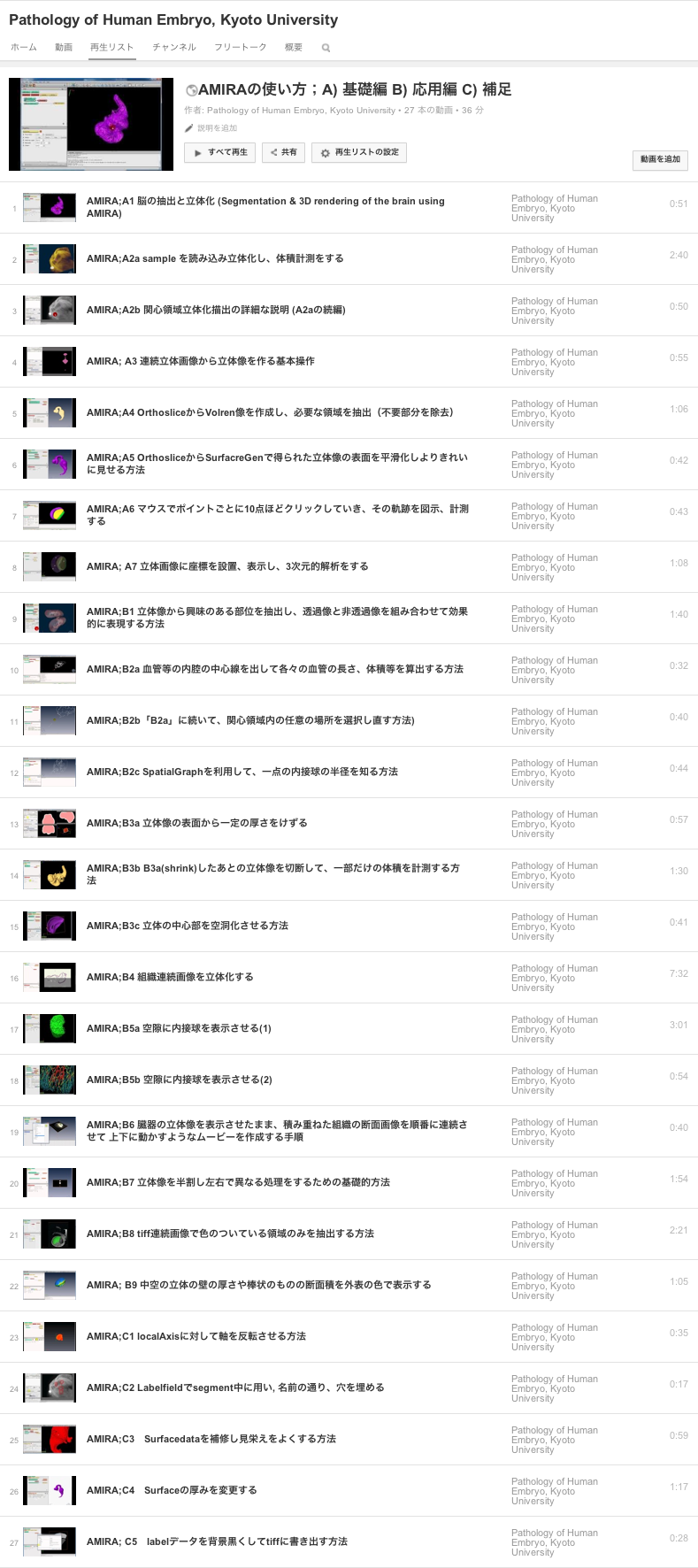
今回、AMIRA等のソフトの使用法を示す動画を整理しました。ご利用ください。(‘14,06,25)  海外君、名古さんの論文で作成した胃の立体像が解剖学雑誌Anatomical Record 297巻5号の表紙に採用されるとともに、ビデオを結果として示すことのできるAR WOW-Video Articleの第1号の論文として採用されました。 N. Kaigai, A. Nako, S. Yamada, C. Uwabe, K. Kose and T. Takakuwa Anat RecはAmerican Association of Anatomists (AAA)の公認雑誌で、100年以上の発行歴のある由緒ある雑誌です。永く世界の医学、解剖学、発生学の分野を牽引してきました。本雑誌は、さらに、最近のe-Page技術を取り入れたWOWという雑誌を本号から採用しました。これは、ビデオが論文の結果として重要な役割を果たす際にそのビデオが永久に保存できるようにしたものです。その第1号の論文として、海外君の胃の形態形成と動きについての論文が選ばれました。 Editorialでは、”Human Embryos are Turning on the e-Pages of AR”(ヒト胚子がARのe-Pageに灯をともす…)という題で, インタビューを受け、私たちの予想以上に讃えていただきました。(Anat Rec 297(5) ; 799–801, 2014) Our rationale for imaging the human stomach during development.“All of the authors are well-versed with the fact that the stomach develops as the local widening of the foregut at Carnegie Stage (CS) 13, as well as the morphology and position of the stomach in adults. But what are the developmental dynamics from the former to the latter? While I (Dr. Takakuwa) was a university student, I read a textbook that explained that the developmental dynamics of the stomach follow the order of linear movement along the caudal direction, rotation around the longitudinal (Z) axis, and rotation around the dorsoventral (X) axis. This explanation aroused my curiosity with regard to the position of the abdominal organs around the stomach, such as the esophagus, pancreas, and duodenum, which are restricted in their positions after CS17. For example, around CS20, movement of the stomach is restricted at both its entrance (cardia) and the exit (pyloric antrum) near the mid-sagittal plane. We designed our study to sort out the dynamic process that places the stomach in its definitive position in the abdomen. Accordingly, we analyzed the external morphology and morphometry of the human embryonic stomach, as well as documented its precise 3D movements, using magnetic resonance (MR) imaging data of human embryos in the “Kyoto Collection”. We discovered that the line connecting the cardia and the pyloric antrum of the stomach does not rotate around the dorsoventral (X) axis, as widely believed, but rotates around the transverse (Y) axis. The stomach “appears” to move towards the left, laterally and caudally, as deflection and differential growth progresses. We found that the developmental morphology of the three-dimensionally reconstructed stomach was not “analogous” to that of adults or as described in recent textbooks. Rather, we found that the stomach’s developmental morphology is as documented in a study a century before (Lewis 1912), in which the stomach was precisely hand drawn by a special artist [note added by Editor: Lewis studied the stomachs of five human embryos that were 10 mm and 45 mm in length; Harvard Embryological Collection, Series 1000]. We are gratified that our MR imaging data of embryos enhance the value of the Kyoto Collection, not only as archives of historical specimens but also as useful research resources for the future.” Contributed by Kaigai and colleagues, Kyoto University, Graduate School of Medicine. 海外くんの卒業論文がAnatomical recordに掲載されました。卒業後、 Office assistantとして2年かけて仕上げてくれました。教科書には、胃の動きは”下降”、”頭尾軸に対する回転”、”背腹軸に対する回転”と3つの動きにわけて説明されています。しかしながら実際の動きは立体空間的でありそう単純ではありませんでした。表紙絵に採用されました。 8. Morphogenesis and three-dimensional movement of the stomach during the human embryonic period, 2014 May;297(5):791-7. doi: 10.1002/ar.22833.
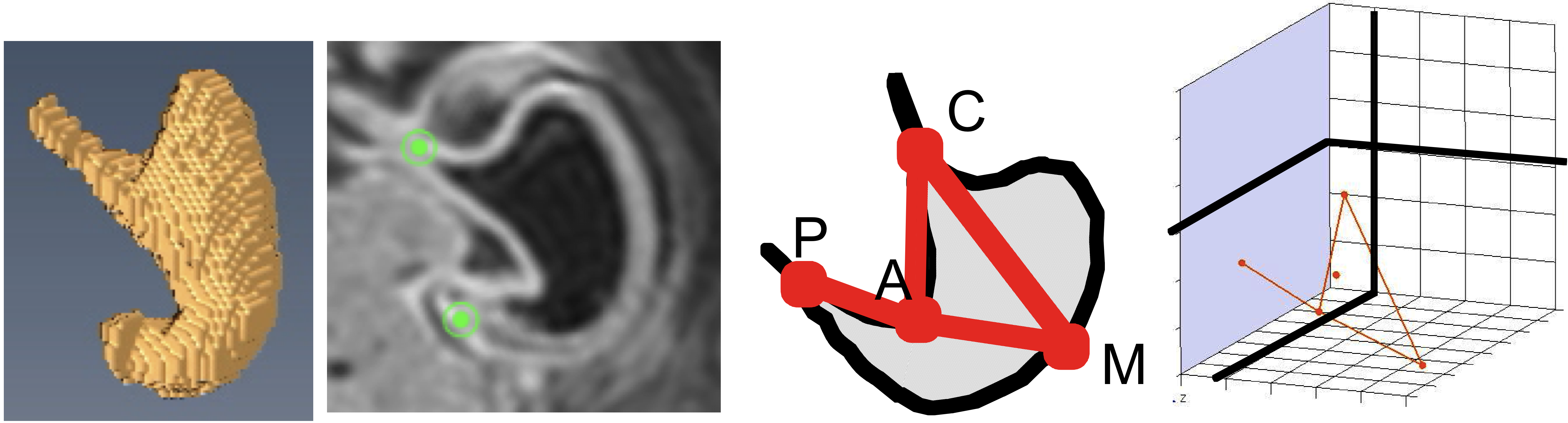 本研究の立体画像元データの一部はMorphoMuseuMに受諾されました。 20. Nako A, Kaigai N, Shiraki N, Yamada S, Uwabe C, Kose K, Takakuwa T, 3D models related to the publication: Morphogenesis of the stomach during the human embryonic period, MorphoMuseuM, in press ABSTRACTThe stomach develops as the local widening of the foregut after Carnegie stage (CS) 13 that moves in a dramatic and dynamic manner during the embryonic period. Using the magnetic resonance images of 377 human embryos, we present the morphology, morphometry, and three-dimensional movement of the stomach during CS16 and CS23. The stomach morphology revealed stage-specific features. The angular incisura and the cardia were formed at CS18. The change in the angular incisura angle was approximately 90° during CS19 and CS20, and was <90° after CS 21. The prominent formations of the fundus and the pylorus differentiate at around CS20. Morphometry of the stomach revealed that the stomach gradually becomes “deflected” during development. The stomach may appear to move to the left laterally and caudally due to its deflection and differential growth. The track of the reference points in the stomach may reflect the visual three-dimensional movement. The movement of point M, representing the movement of the greater curvature, was different from that of points C (cardia) and P (pyloric antrum). The P and C were located just around the midsagittal plane in all the stages observed. Point M moved in the caudal-left lateral direction until CS22. Moreover, the vector CP does not rotate around the dorsoventral axis, as widely believed, but around the transverse axis. The plane CPM rotated mainly around the longitudinal axis. The data obtained will be useful for prenatal diagnosis in the near future. |
|||