アクセスカウンター

2020年9月
 123456
78910111213
14151617181920
21222324252627
282930  

西谷さん、鳥居さんの胎児心臓の解析がJAHAに掲載

西谷さんの修士論文、鳥居さんの卒業論文;胎児心臓のDT-MRIによる解析がJAHAに掲載されました。

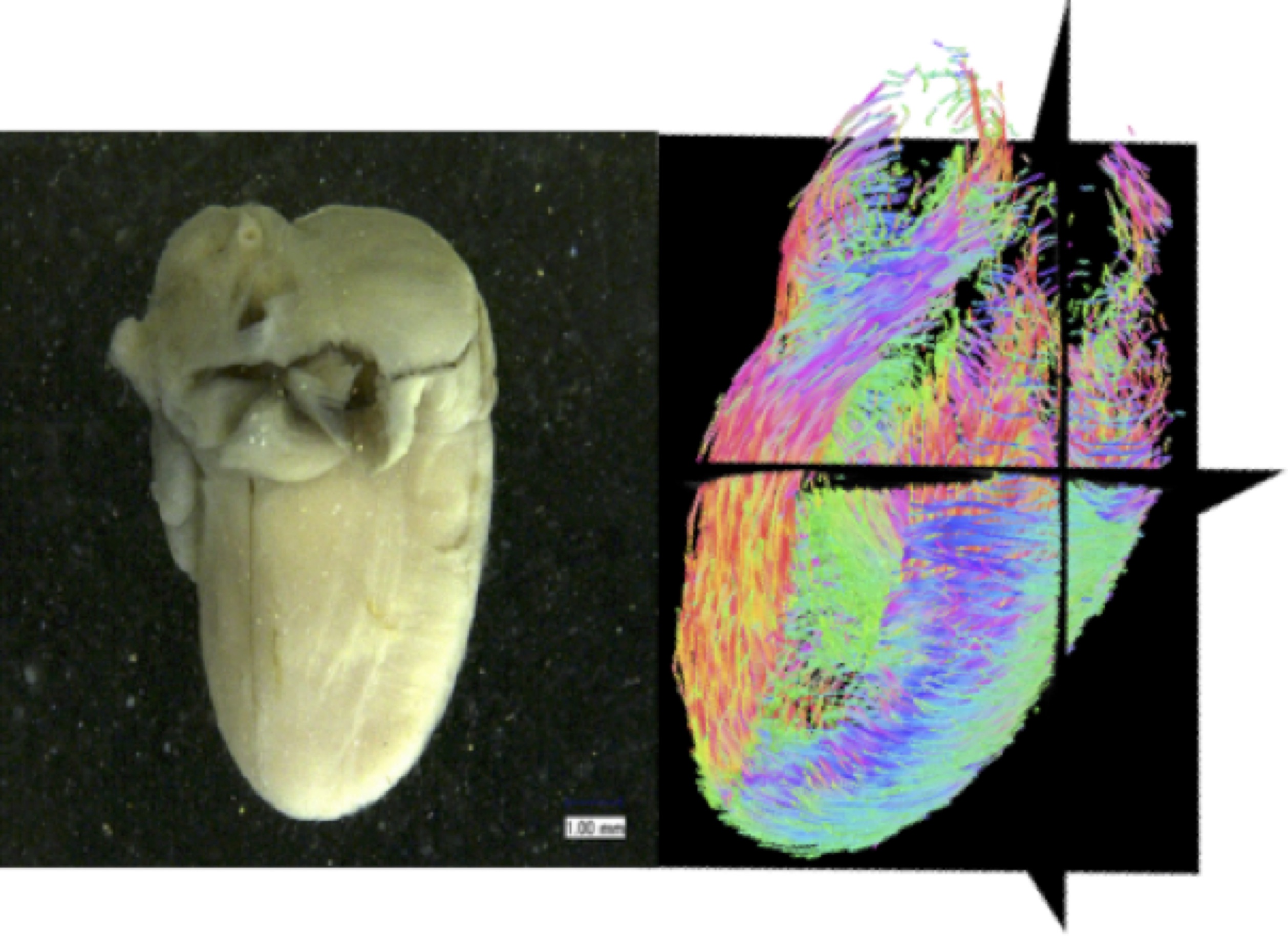
  • 従来の報告より8週以上若い時期のヒトの心筋走行を高解像度DT-MRIデータを用いて検出しました。
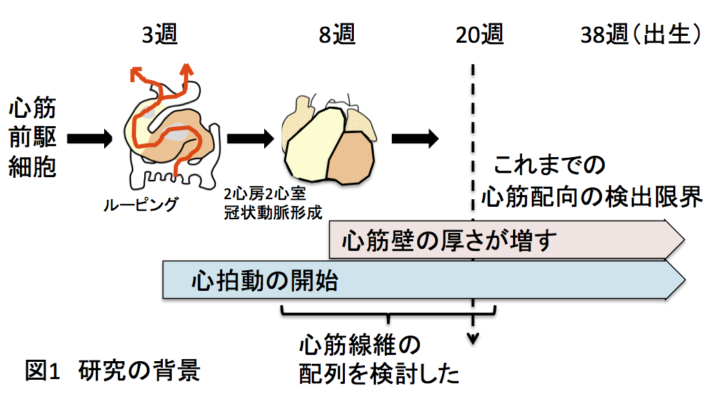
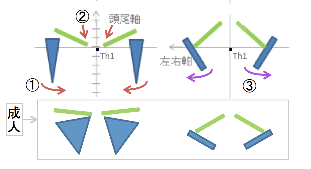
  • ヒトの心筋の走行の配置、螺旋状の角度等は、成人とすでに同様であることから、心筋構築の青写真は胚子期末(CS20-23, GSA8W)には、すでに形成されていることをから明らかにしました。
  • データは古典的な心筋バンド説(心筋はひとつながりのバンド状のものが立体的に折り畳まれて形成されている)を否定しました。

本研究成果は、京都大学HP(トピックス)でも紹介されました。

ヒト胎児心臓の心筋線維方向を追跡 -受精後8週の心筋線維は成人と同じ配列をする-  外部リンク[京都大学HP(トピックス)]

2020/10/13 医療NEWSにて,ヒト胎児の心臓、受精後8週で心筋線維の配列が成人と同様であることが判明-京大ほか が紹介されました.

46. Nishitani S, Torii N, Imai H, Haraguchi R, Yamada S, Takakuwa T, Development of helical myofiber tracts in the human fetal heart: Analysis of myocardial fiber formation in the left ventricle from the late human embryonic period using diffusion tensor magnetic resonance imaging. Journal of the American Heart Association, 2020, 19(9) doi:10.1161/JAHA.120.016422

Abstract

Background

Detection of the fiber orientation pattern of the myocardium using diffusion tensor magnetic resonance imaging lags ≈12 weeks of gestational age (WGA) behind fetal myocardial remodeling with invasion by the developing coronary vasculature (8 WGA). We aimed to use diffusion tensor magnetic resonance imaging tractography to characterize the evolution of fiber architecture in the developing human heart from the later embryonic period.

Methods and Results

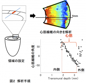
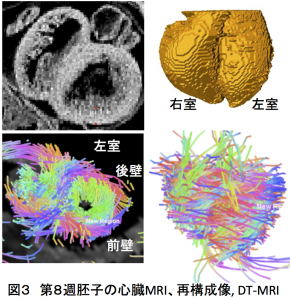
Twenty human specimens (8–24 WGA) from the Kyoto Collection of Human Embryos and Fetuses, including specimens from the embryonic period (Carnegie stages 20–23), were used. Diffusion tensor magnetic resonance imaging data were acquired with a 7T magnetic resonance system. Fractional anisotropy and helix angle were calculated using standard definitions. In all samples, the fibers ran helically in an organized pattern in both the left and right ventricles. A smooth transmural change in helix angle values (from positive to negative) was detected in all 16 directions of the ventricles. This feature was observed in almost all small (Carnegie stage 23) and large samples. A higher fractional anisotropy value was detected at the outer side of the anterior wall and septum at Carnegie stage 20 to 22, which spread around the ventricular wall at Carnegie stage 23 and in the early fetal samples (11–12 WGA). The fractional anisotropy value of the left ventricular walls decreased in samples with ≥13 WGA, which remained low (≈0.09) in larger samples.

Conclusions

From the human late embryonic period (from 8 WGA), the helix angle arrangement of the myocardium is comparable to that of the adult, indicating that the myocardial structure blueprint, organization, and integrity are already formed.

ヒト胎児心臓の心筋線維方向の追跡 ―受精後8週の心筋線維は成人と同じ配列をする―

概要

心臓は受精後3週頃には拍動を開始し生涯拍動を続けます。全身に効率よく血液を送るために心筋線維は立体的に規則的に配列される必要があります。近年、拡散テンソルMRIを用いて、心筋線維がどのように配列されているかについても検討できるようになりました。成人では心室の壁を構成する心筋線維は、層状に内側から外側にかけてゆるやかに向きを変えて配列することが示されています。胎児では、胎齢20週ころには成人と同様の心筋線維配列を示すことが知られていますが、拍動開始から20週の間の心筋線維の配列は不明でした。われわれは、京都大学大学院医学研究科附属先天異常標本解析センターが所蔵しているヒト胚標本20体を用いて、ヒト心臓の心筋線維の配列を拡散テンソルMRIを用いて検討しました。その結果、心室の壁が厚みをもち始める胚子期後半の段階において、心筋線維の配列は、成人と同様であること証明しました。今回の結果は、先天性、後天性心疾患の原因解明、診断、治療等に関わる重要な知見と考えられます。

1.背景

心臓は受精後3週頃には拍動を開始し生涯拍動を続ける、生きるために必須の臓器で、成人では200-300gあります。心臓の大部分は心筋という特殊な筋肉から作られています。特に心室は心筋が袋状になり、全身に効率よく血液を送るために心筋線維が規則的に配列される必要があります。心筋線維の配列は、解剖学者Torrent-Guaspの心筋バンド説(心臓の壁を構成する心筋を広げていくと、1枚の帯状のものになる)が長い間信じられていました。近年、MRIという機器を用いて、心臓を取り出さずに、その形態や構成する物質の性質を調べることが可能になり、心筋線維がどのように配列されているかについても拡散テンソルMRI(DT-MRI)法という解析方法を用いて検討できるようになりました。心室の壁を構成する心筋線維の向きは、どの壁においても、層状に内側から外側にかけてゆるやかに向きを変え、らせん状に配列することが示されており(メッシュモデル)、心筋バンド説は否定されています。

ヒトに心臓の心筋線維の配列は、いつごろからどのように形成されるのでしょうか。これまでの研究では、胎齢20週ころにようやく成人と同様の配列を示すことが報告されています。そうすると、拍動開始(3週)から20週の間も、心臓は拍動しているわけですが、その間、心筋の配列はどうなっているのかという疑問が湧いてきます。そこで我々は、ヒト心臓の心筋線維の配列を24週の胎児から発生をさかのぼって検討することにしました。(図1)

2.研究手法・成果

心筋の壁が組織学的に形成されるには、心臓が2心房2心室の4つの部屋に別れ、冠状動脈という血管が形成され、心筋への血流が確保されるカーネギーステージ(CS)22(第8週中頃)であることがわかっています。そこで、それよりも若いCS20(第8週初め)までを解析対象としました。

解析の成功のためには、良質なヒト胚子標本の確保と、小さい標本を解析するために解像度の高い撮像装置、撮像条件の検討、解析法の確立が重要です。(胚子期後半の心臓は米粒ほどの大きさしかありません!!)

良質なヒト胚子標本として、附属先天異常標本解析センターが収集、保管している、ヒト胎児大規模標本群(京都コレクション)を使用しました。この標本群は、世界最大規模の研究リソースとして知られており、その利用については京都大学大学院医学研究科・医学部及び医学部附属病院 医の倫理委員会の承認のもとで研究が行われています。撮像装置としては、医学研究科医学研究支援センターが保有している前臨床用7T-MRIを用いました。撮像の対象及び目的に特化させた撮像条件と直径19mmの高感度MRIコイルを併用してDTI撮像データの質の改善を行いました。

その結果、心室の壁が形成され厚みをもち始める胚子期後半の段階(CS20-23)においても、心筋線維の配列は、成人と同様の配列を示すことを示すことができました。(図2、3)

3.波及効果、今後の予定

心臓の病気には、発生過程の異常に基づくものが多くあります。その意味で、今回心臓の動力の源である心筋の配列過程を経時的に追求できたことは大きな意義があります。ただ、心拍動の開始から8週までの期間については、装置の解像度の問題から現在の方法での解析が難しく今後の課題と言えます。今回の結果は、先天性、後天性心疾患の原因解明、診断、治療等に関わる重要な知見で、その発展に貢献することが期待されます。また、今回得られたDT-MRI撮像、解析技術は、心臓以外の他の臓器の形態形成の解析にも応用できるものと考えられます。

<研究者のコメント>

胚子期後半の心筋線維の配列がすでに大人の配列と同様というのは、当然とは言い切れません。発生学の原則として、“ヒト胚子はその発生の各段階で生きており、各段階で適切な状態を維持している”(Heuser & Streeter 1941)があります。それに従えば、心臓(心筋)は、成人の完成図を目指して性急な成長をするのではなく、発生段階の心臓の形状や他の諸器官と協調して、心筋の走行や分布、成長が異なることも十分に想定されることだからです。

<西谷さんの修士論文の概要>

Nishitani S, Torii N, Imai H, Haraguchi R, Yamada S, Takakuwa T, Development of helical myofiber tracts in the human fetal heart: Analysis of myocardial fiber formation in the left ventricle from the late human embryonic period using diffusion tensor magnetic resonance imaging. Journal of the American Heart Association, 2020,  19(9) doi:10.1161/JAHA.120.016422

田中さん、坂本さんの卒業研究がPLoS ONEに掲載

田中さん、坂本さんの卒業研究がPLoS ONEに掲載されました。胎児期の上肢帯の形態形成、位置の変化について詳述しました。

  • CS18で、烏口骨と上腕骨頭とともに肩甲骨体部を最初に検出
  • 肩甲骨下角は尾側方向に拡大したが、頭側はほとんど位置をかえない
  • 肩甲骨は胎児期には独特な位置(椎体の前方から側方への移動)、向き( 内部、上方回転)を示す

46. Tanaka S, Sakamoto R, Kanahashi T, Yamada S, Imai H, Yoneyama A, Takakuwa T. Shoulder girdle formation and positioning during embryonic and early fetal human development. PLoS ONE 2020, 15(9): e0238225. https://doi.org/10.1371/journal.pone.0238225

Abstract

Positional information on the shoulder girdle (the clavicle and scapula) is important for a better understanding of the function of the upper limb in the locomotive system as well as its associated disease pathogenesis. However, such data are limited except for information on the axial position of the scapula. Here, we describe a three-dimensional reconstruction of the shoulder girdle including the clavicle and scapula, and its relationship to different landmarks in the body. Thirty-six human fetal specimens (crown-rump length range: 7.6–225 mm) from the Kyoto Collection were used for this study. The morphogenesis and three-dimensional position of the shoulder girdle were analyzed with phase-contrast X-ray computed tomography and magnetic resonance imaging. We first detected the scapula body along with the coracoid and humeral head at Carnegie stage 18; however, the connection between the body and coracoid was not confirmed at this stage. During development, all landmarks on the shoulder girdle remained at the same axial position except for the inferior angle, which implies that the scapula enlarged in the caudal direction and reached the adult axial position in the fetal period. The scapula body was rotated internally and in the upward direction at the initiation of morphogenesis, but in the fetal period the scapula body was different than that in the adult position. The shoulder girdle was located at the ventral side of the vertebrae at the time of initial morphogenesis, but changed its position to the lateral side of the vertebrae in the late embryonic and fetal periods. Such a unique position of the shoulder girdle may contribute to the stage-specific posture of the upper limb. Adequate internal and upward rotation of the scapula could help in reducing the shoulder width, thereby facilitating childbirth. The data presented in this study can be used as normal morphometric references for shoulder girdle evaluations in the embryonic and fetal periods.