Fukui N, Toru KanahashiT, MatsubayashiJ, ImaiH, YoneyamaA, OtaniH, YamadaS, Takakuwa T. Morphogenesis of the pulmonary vein and left atrial appendage in human embryos and early fetuses. J Anatomy 2024, 244, 142-158, in press, https://doi.org/10.1111/joa.13941
Abstract
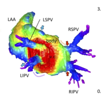
The left atrium wall has several origins, including the body, appendage, septum, atrial–ventricular canal, posterior wall, and venous component. Here, we describe the morphogenesis of left atrium based on high-resolution imaging (phase-contrast X-ray computed tomography and magnetic resonance imaging). Twenty-three human embryos and 19 fetuses were selected for this study. Three-dimensional cardiac images were reconstructed, and the pulmonary veins and left atrium, including the left atrial appendage, were evaluated morphologically and quantitatively. The positions of the pericardial reflections were used as landmarks for the border of the pericardial cavity. The common pulmonary vein was observed in three specimens at Carnegie stage 17–18. The pericardium was detected at the four pulmonary veins (left superior, left inferior, right superior, and right inferior pulmonary veins) at one specimen at Carnegie stage 18 and all larger specimens, except the four samples. Our results suggest that the position of the pericardial reflections was determined at two pulmonary veins (right and left pulmonary vein) and four pulmonary veins almost simultaneously when the dorsal mesocardial connection between the embryo and heart regressed. The magnetic resonance images and reconstructed heart cavity images confirmed that the left atrium folds were present at the junction between the body and venous component. Three-dimensional reconstruction showed that the four pulmonary veins entered the dorsal left atrium tangentially from the lateral to the medial direction. More specifically, the right pulmonary veins entered at a greater angle than the left pulmonary veins. The distance between the superior and inferior pulmonary veins was shorter than that between the left and right pulmonary veins. Three-dimensional reconstruction showed that the venous component increased proportionally with growth. No noticeable differences to discriminate between the right and left parts of the venous component emerged, while the junction between the venous component and body gradually became inconspicuous but was still recognizable by the end of the observed early fetal period. The left superior pulmonary vein had the smallest cross-sectional area and most flattened shape, whereas the other three were similar in area and shape. The left atrial appendage had a large volume in the center and extended to the periphery as a lobe-like structure. The left atrial appendage orifice increased in the area and tended to become flatter with growth. The whole left atrium volume^(1/3) increased almost proportionally with growth, parallel to the whole heart volume. This study provided a three-dimensional and quantitative description of the developmental process of left atrium, comprising the venous component and left atrial appendage formation, from the late embryonic to the early fetal stages.
60. Takakuwa T, Saizonou MA, Fujii S, Kumano Y, Ishikawa A, Aoyama T, Imai H, Yamada S, Kanahashi T. Femoral posture during embryonic and early fetal development: An analysis using landmarks on the cartilaginous skeletons of ex vivo human specimens. PLOS one, 2023, 18(5): e0285190. https://doi.org/10.1371/journal.pone.0285190.
Abstract
The pre-axial border medially moves between the fetal and early postnatal periods, and the foot sole can be placed on the ground. Nonetheless, the precise timeline when this posture is achieved remains poorly understood. The hip joint is the most freely movable joint in the lower limbs and largely determines the lower-limb posture. The present study aimed to establish a timeline of lower-limb development using a precise measurement of femoral posture. Magnetic resonance images of 157 human embryonic samples (Carnegie stages [CS] 19–23) and 18 fetal samples (crown rump length: 37.2–225 mm) from the Kyoto Collection were obtained. Three-dimensional coordinates of eight selected landmarks in the lower limbs and pelvis were used to calculate the femoral posture. Hip flexion was approximately 14° at CS19 and gradually increased to approximately 65° at CS23; the flexion angle ranged from 90° to 120° during the fetal period. Hip joint abduction was approximately 78° at CS19 and gradually decreased to approximately 27° at CS23; the average angle was approximately 13° during the fetal period. Lateral rotation was greater than 90° at CS19 and CS21 and decreased to approximately 65° at CS23; the average angle was approximately 43° during the fetal period. During the embryonic period, three posture parameters (namely, flexion, abduction, and lateral rotation of the hip) were linearly correlated with each other, suggesting that the femoral posture at each stage was three-dimensionally constant and exhibited gradual and smooth change according to growth. During the fetal period, these parameters varied among individuals, with no obvious trend. Our study has merits in that lengths and angles were measured on anatomical landmarks of the skeletal system. Our obtained data may contribute to understanding development from anatomical aspects and provide valuable insights for clinical application.
59. Matsunari C, Kanahashi T, Otani H, Imai H, Yamada S, Okada T, Takakuwa T. Tentorium cerebelli formation during human embryonic and early fetal development. Anat Rec (Hoboken) 2023, 306(3), 515-526
Abstract
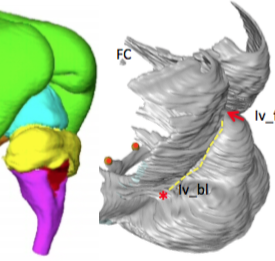
The morphologies of the fetal tentorium cerebelli (TC) and brain influence each other during development. This study aimed to analyze and more comprehensively understand the three-dimensional morphogenesis of the TC and fetal brain. We examined magnetic resonance imaging from 64 embryonic and fetal specimens (crown-rump length range, 9.2–225 mm). During the embryonic period, the lateral folds of the TC elongated to traverse the middle part of the midbrain. The TC and falx cerebri appeared separated, and no invaginations at the parieto-occipital region were observed. In the early fetal period, the cerebrum covered approximately half of the midbrain. The separation of the dural limiting layer at the parieto-occipital region widened from the posterior cerebrum to the cranial cerebellum. The lateral folds of the TC were spread between its tip, continuous with the falx cerebri, and its base plane, located between the midbrain and rostral hindbrain. Differences in the TC components’ growth directions gradually diminished as the cerebrum covered the midbrain. We observed rotation of the TC at its median section according to its growth, which ceased in the middle fetal period. The brainstem and cerebellum extended inferiorly via differential growth, with the cerebrum covering them superiorly. The morphology of the TC curved to conform to the cerebellar and cerebral surfaces. Our present study suggests that factors affecting TC morphology differ between the early and middle fetal periods. Present data provided a more comprehensive view of TC formation according to developmental stage.
Kanahashi T, Imai H, Otani H, Yamada S, Yoneyama A, Takakuwa T. Three-dimensional morphogenesis of the human diaphragm during the late embryonic and early fetal period: Analysis using T1-weighted and diffusion tensor imaging. J Anat. 2023, 242, 174-190, DOI: 10.1111/joa.13760
Abstract
表紙に採用されました
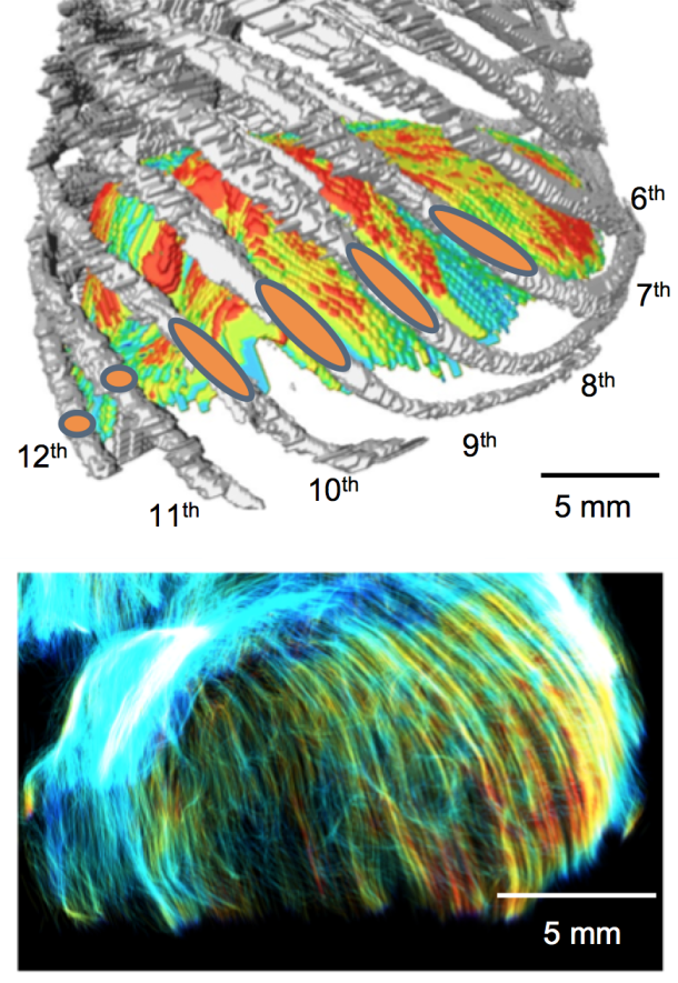
A precise understanding of human diaphragm development is essential in fetal medicine. To our knowledge, no previous study has attempted a three-dimensional (3-D) analysis and evaluation of diaphragmatic morphogenesis and development from the embryonic to the early fetal period. This study aimed to evaluate the morphogenesis and fibrous architecture of the developing human diaphragm during the late embryonic and early fetal periods. Fifty-seven human embryos and fetuses (crown-rump length [CRL] = 8–88 mm) preserved at the Congenital Anomaly Research Center of Kyoto University and Shimane University were analyzed. 3-D morphogenesis and fiber orientation of the diaphragm were assessed using phase-contrast X-ray computed tomography, T1-weighted magnetic resonance imaging (T1W MRI), and diffusion tensor imaging (DTI). T1W MR images and DTI scans were obtained using a 7-T MR system. The diaphragm was completely closed at Carnegie stage (CS) 20 and gradually developed a dome-like shape. The diaphragm was already in contact with the heart and liver ventrally in the earliest CS16 specimen observed, and the adrenal glands dorsally at CS19 or later. In the fetal period, the diaphragm contacted the gastric fundus in samples with a CRL ≥41 mm, and the spleen in samples with a CRL ≥70 mm. The relative position of the diaphragm with reference to the vertebrae changed rapidly from CS16 to CS19. The most cranial point of the diaphragm was located between the 4th and 8th thoracic vertebrae, regardless of fetal growth, in samples with a CRL ≥16 mm. Diaphragmatic thickness was nearly uniform (0.15–0.2 mm) across samples with a CRL of 8 mm to 41 mm. The sternal, costal, lumbar parts, and the area surrounding the esophageal hiatus thickened with growth in samples with a CRL ≥46 mm. The thickness at the center of the diaphragm and the left and right hemidiaphragmatic domes did not increase with growth. Tractography showed that the fiber orientation of the sternal, costal, and lumbar parts became more distinct as growth progressed in CS19 or later. All fibers in the costal and lumbar regions ran toward the left and right hemidiaphragmatic domes, except for those running to the caval opening and esophageal hiatus. The fiber orientation patterns from the right and left crura surrounding the esophageal hiatus were classified into three types. Distinct fiber directions between the costal and sternal, and between the costal and lumbar diaphragmatic parts were observable in samples with a CRL ≥46 mm. Anterior costal and sternal fibers ran toward the center. Fiber tracts around the center and the left and right hemidiaphragmatic domes; between the costal and lumbar orientations; and between the costal and sternal orientations showed a tendency for decreasing fractional anisotropy values with fetal growth, and showed less density than other areas. In conclusion, we used 3-D thickness assessment and DTI tractography to identify qualitative changes in the muscular and tendonous regions of the diaphragm during the embryonic and early fetal periods. This study provides information on normal human diaphragm development for the progression of fetal medicine and furthering the understanding of congenital anomalies.
Nohara A, Owaki N, Matsubayashi J, Katsube M, Imai H, Yoneyama A, Yamada S, Kanahashi T, Takakuwa T. Morphometric analysis of secondary palate development in human embryos. J Anatomy, 2022, 241(6), 1287-1302, 2022, DOI:10.1111/joa.13745
Abstract
Rapid shelf elevation and contact of the secondary palate and fusion reportedly occur due to a growth-related equilibrium change in the structures within the oro-nasal cavity. This study aimed to quantitatively evaluate complex three-dimensional morphological changes and their effects on rapid movements, such as shelf elevation and contact, and fusion. Morphological changes during secondary palate formation were analyzed using high-resolution digitalized imaging data (phase-contrast X-ray computed tomography and magnetic resonance images) obtained from 22 human embryonic and fetal samples. The three-dimensional images of the oro-nasal structures, including the maxilla, palate, pterygoid hamulus, tongue, Meckel’s cartilage, nasal cavity, pharyngeal cavity, and nasal septum, were reconstructed manually.
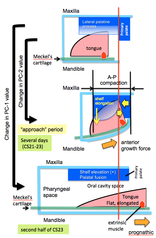
palatal shelves were not elevated in all the samples at Carnegie stage (CS)21 and CS22 and in three samples at CS23. In contrast, the palatal shelves were elevated but not in contact in one sample at CS23. Further, the palatal shelves were elevated and fused in the remaining four samples at CS23 and all three samples from the early fetal period. For each sample, 70 landmarks were subjected to Procrustes and principal component (PC) analysis. PC-1 accounted for 67.4% of the extracted gross changes before and after shelf elevations. Notably, the PC-1 values of the negative and positive value groups differed significantly. The PC-2 value changed during the phases in which the change in the PC-1 value was unnaturally slow and stopped at CS22 and the first half of CS23. This period, defined as the “approach period”, corresponds to the time before dynamic changes occur as the palatal shelves elevate, the tongue and mandibular tip change their position and shape, and secondary palatal shelves contact and fuse. During the “approach period”, measurements of PC-2 changes showed that structures on the mandible (Meckel’s cartilage and tongue) and maxilla (palate and nasal cavity) did not change positions, albeit both groups of structures appeared to be compressed anterior-posteriorly. However, during and after shelf elevation, measurements of PC-1 changes showed significant changes between maxillary and mandibular structures, particularly positioning of the shelves above the tongue and protrusion of the tongue and mandible. These results suggest an active role for Meckel’s cartilage growth in repositioning the tongue to facilitate shelf elevation. The present data representing three distinct phases of secondary palate closure in humans can advance the understanding of morphological growth changes occurring before and after the horizontal positioning of palatal shelves and their fusion to close the secondary palate in humans successfully.
KakeyaM, Matsubayashi J, Kanahashi T, Männer J, Yamada S, Takakuwa T. The return process of physiological umbilical herniation in human fetuses: the possible role of the vascular tree and umbilical ring. J Anatomy 2022, 241(3), 846-859. https://doi.org/10.1111/joa.13720
Abstract
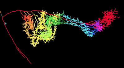
The human intestine elongates during the early fetal period, herniates into the extraembryonic coelom (EC), and subsequently returns to the abdominal cavity (AC). The process by which the intestinal loop returns to the abdomen remains unclear. This study aimed to document positional changes in the intestinal tract with the superior mesenteric artery (SMA) and branches in 3D to elucidate the intestinal loop return process (transition phase). Serial histological cross-sections from human fetuses (crown–rump length [CRL] range: 30–50 mm) in the herniation (n = 1), transition (n = 7), and return (n = 2) phases were selected from the Blechschmidt Collection. The distribution of the SMA trunk and all intestinal and sister branches entering the intestines was visualized so that positional changes in branches were continuous from the herniation to return phases. Positional changes in SMA branches proceeded in an orderly and structured manner; this is essential for continuous blood supply via the SMA to the intestine during transition and for safe intestinal return. Changes in the SMA distribution proceeded prior to the detection of initiation of intestinal tract return, which might start earlier and last much longer than our consensus (i.e., that the return of the herniated intestine begins when the CRL is approximately 40 mm and ends within a short time). In the cross-section of the umbilical ring in the herniation and transition phases, one proximal limb and one distal limb were observed with SMA intestinal branches, which were fully packed in the umbilical ring. The SMA branches were aligned from inferior to superior along the SMA main trunk. In the herniation phase, the distribution of 3rd–13th branches aligned from proximal inferior medial to distal superior left with a slight spiral in the EC, the tips of which suggested an orderly running course of the small intestine. In the transition phase, SMA branches running across the umbilical ring that fed the small intestine were observed, suggesting that the intestine was uncoiled and ran across the umbilical ring almost vertically. The estimated curvature value supported the phenomenon of uncoiling at the umbilical ring; the value at the umbilical ring was lesser than that in the AC and EC. During the transition phase, the proximal and distal limbs transversely ran side by side in the AC, umbilical ring, limbs on the cranial side, and mesentery on the caudal side. The SMA trunk and its branches ran in parallel, cranially to caudally aligned in the mesentery. This layout of the umbilical ring was maintained during the transition phase. In the return phase, the SMA trunk was gently curved from the upper left to the lower right of the AC; around 12 branches spread with a winding staircase appearance. The intestinal tract reached its definitive position immediately after all tissues crossed the umbilical ring and released any restriction. Each SMA branch and the corresponding region of the intestinal tract form a unit and change their position, though the conformation may change within each unit when running across the umbilical ring. We suggest that the slide–stack model requires revision.
Kumano Y, Tanaka S, Sakamoto R, Kanahashi T, Imai H, Yoneyama A, Yamada S, Takakuwa T. Upper arm posture during human embryonic and fetal development. Anatomical Rec 2022, 305 1682-1691, https://doi.org/10.1002/ar.24796
Abstract
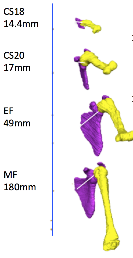
The upper extremity posture is characteristic of each Carnegie stage (CS), particularly between CS18 and CS23. Morphogenesis of the shoulder joint complex largely contributes to posture, although the exact position of the shoulder joints has not been described. In the present study, the position of the upper arm was first quantitatively measured, and the contribution of the position of the shoulder girdle, including the scapula and glenohumeral (GH) joint, was then evaluated. Twenty-nine human fetal specimens from the Kyoto Collection were used in this study. The morphogenesis and three-dimensional position of the shoulder girdle and humerus were analyzed using phase-contrast X-ray computed tomography and magnetic resonance imaging. Both abduction and flexion of the upper arm displayed a local maximum at CS20. Abduction gradually decreased until the middle fetal period, which was a prominent feature. Flexion was less than 90° at the local maximum, which was discrepant between appearance and measurement value in our study. The scapular body exhibited a unique position, being oriented internally and in the upward direction, with the glenoid cavity oriented cranially and ventrally. However, this unique scapular position had little effect on the upper arm posture because the angle of the scapula on the thorax was canceled as the angle of the GH joint had changed to a mirror image of that angle. Our present study suggested that measuring the angle of the scapula on the thorax and that of the GH joint using sonography leads to improved staging of the human embryo.
54. Yamazaki Y, Kanahashi T, Yamada S, Männer J, Takakuwa T. Three-dimensional analysis of human laryngeal and tracheobronchial cartilages during the late embryonic and early fetal period. Cells Tissues Organs, 2021 in press
Abstract
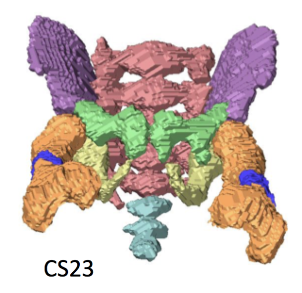
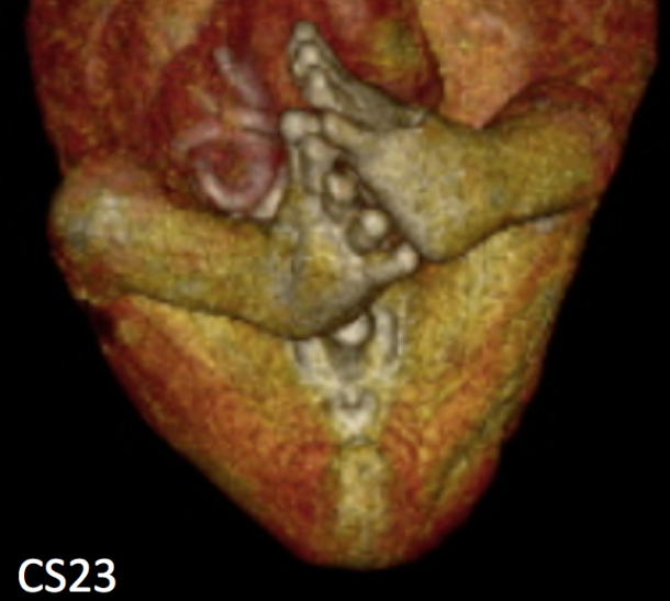
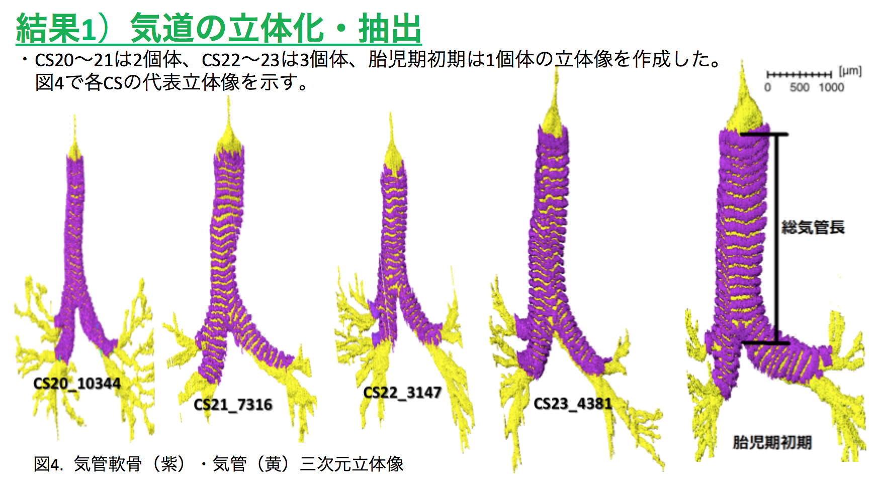
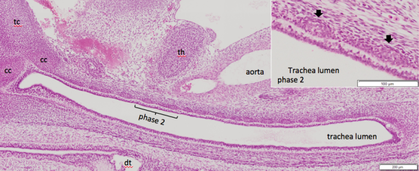
Laryngeal and tracheobronchial cartilages are present as unique U-shaped forms around the respiratory tract and contribute to the formation of rigid structures required for the airway. Certain discrepancies still exist concerning cartilage formation in humans. To visualize the accurate timeline of cartilage formation, tracheobronchial and laryngeal cartilages were 3D reconstructed based on serial tissue sections during the embryonic period (Carnegie stage [CS] 18–23) and early fetal period (crown rump length [CRL] = 35–45 mm). The developmental phases of the cartilage were estimated by histological studies, which were performed on the reconstructed tissue sections. The hyoid greater horns were recognizable at CS18 (phase 2). Fusion of 2 chondrification centers in the mid-sagittal region was observed at CS19 in the hyoid bone, at CS20 in the cricoid cartilage, and in the specimen with CRL 39 mm in the thyroid cartilage. Phase 3 differentiation was observed at the median part of the hyoid body at CS19, which was the earliest among all other laryngeal and tracheobronchial cartilages. Most of the laryngeal cartilages were in phase 3 differentiation at CS22 and in phase 4 differentiation at CS23. The U-shaped tracheobronchial cartilages with phase 2 differentiation covered the entire extrapulmonary region at CS20. Phase 3 differentiation started on the median section and propagates laterally at CS21. The tracheobronchial cartilages may form simultaneously during the embryonic period at CS22-23 and early fetal periods, similar to adults in number and distribution. The spatial propagation of the tracheal cartilage differentiation provided in the present study indicates that cartilage differentiation may have propagated differently on phase 2 and phase 3. This study demonstrates a comprehensible timeline of cartilage formation. Such detailed information of the timeline of cartilage formation would be useful to improve our understanding of the development and pathophysiology of congenital airway anomalies.